Giardiavirus internal ribosome entry site has an apparently unique mechanism of initiating translation

8Citations
Citations of this article
37Readers
Mendeley users who have this article in their library.

Abstract

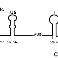
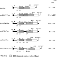
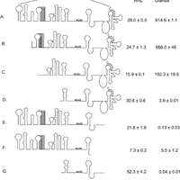
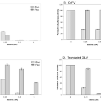
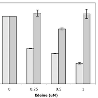
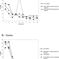
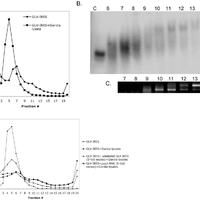
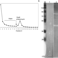
Giardiavirus (GLV) utilizes an internal ribosome entry site (IRES) for translation initiation in the early branching eukaryote Giardia lamblia. Unlike most of the viral IRESs among higher eukaryotes, which localize primarily within the 5′-untranslated region (UTR), the GLV IRES comprises 253 nts of 5′UTR and the initial 264 nts in the open-reading-frame (ORF). To test if GLV IRES also functions in higher eukaryotic systems, we examined it in rabbit reticulocyte lysate (RRL) and found that it functions much less efficiently than the IRES from the Encephalomyocarditis virus (EMCV) or Cricket paralysis virus (CrPV). In contrast, both EMCV-IRES and CrPV-IRESs were inactive in transfected Giardia cells. Structure-function analysis indicated that only the stem-loop U5 from the 5′UTR and the stem-loop I plus the downstream box (Dbox) from the ORF of GLV IRES are required for limited IRES function in RRL. Edeine, a translation initiation inhibitor, did not significantly affect the function of GLV IRES in either RRL or Giardia, indicating that a pre-initiation complex is not required for GLV IRES - mediated translation initiation. However, the small ribosomal subunit purified from Giardia did not bind to GLV IRES, indicating that additional protein factors may be necessary. A member of the helicase family IBP1 and two known viral IRES binding proteins La autoantigen and SRp20 have been identified in Giardia that bind to GLV IRES in vitro. These three proteins could be involved in facilitating small ribosome recruitment for initiating translation. © 2009 Garlapati, Wang.

Figures

  • Figure 1. The essential secondary structures of GLV IRES. Secondary structures identified in the GLV IRES by chemical/enzymatic structure probing and site-directed mutagenesis. doi:10.1371/journal.pone.0007435.g001
  • Figure 2. GLV IRES activity in RRL and in transfected Giardia WB trophozoites. Transcripts were synthesized from various dicistronic cDNA constructs each consisting of the Renilla luciferase Rluc (shaded box) and Photinus luciferase Fluc (hatched box) genes flanked by the 59 and 39 portions of GLV cDNA (the coding region, dash-hatched box; 59 and 39 UTRs, black bars) and located downstream from the T7 promoter (black box). The Rluc and Fluc cistrons are separated by 10 nucleotides in the control pC631Rluc-Fluc transcript (indicated by small black box). Schematic diagrams of dicistronic cDNA constructs (1) pC631Rluc-Fluc, (2) pC631Rluc-UTR-Fluc, (3) pC631Rluc-Cod-Fluc, (4) pC631Rluc-UTRCod-Fluc, (5) pC631Rluc-EMCV-Fluc, and (6) pC631Rluc-CrPV-Fluc are presented. Reaction products from RRL and transfected Giardia trophozoites were assayed for Ruc and Fluc activities and the IRES activity observed with each transcript was expressed as a ratio between the two luciferase activities (Fluc/Rluc 6103). doi:10.1371/journal.pone.0007435.g002
  • Figure 3. Structural requirements for GLV IRES activity in RRL and in Giardia. Effect of varying deletions in the 59UTR and in the 264 nt coding region on GLV-IRES activity observed in RRL and in transfected G. lamblia WB trophozoites. The extent of deletion in each region is mentioned in the text and referred by the number of the transcript in the figure. The Rluc and Fluc activities were assayed from RRL reaction mixture and Giardia lysates. ND, not determined. doi:10.1371/journal.pone.0007435.g003
  • Figure 4. Effect of edeine on IRES functions in RRL. The translation of dicistronic transcripts (A) Cap-Rluc-EMCV IRES-Fluc, (B) Cap-Rluc-CrPV IRES-Fluc, (C) Cap-Rluc-GLV IRES (1–631 nts) Fluc and (D) Cap-Rluc-GLV IRES (276–487) Fluc in RRL, in the presence of varying concentrations of edeine was assayed. The luciferase activity of Rluc (dotted bars) and Fluc (gray bars) are presented as percentage (%) relative luciferase activities as compared to the untreated control samples (without edeine). Error bars represent standard errors from assaying triplicate samples in the same experiment. doi:10.1371/journal.pone.0007435.g004
  • Figure 5. Effect of edeine on the GLV IRES activity in transfected G. lamblia WB trophozoites. The dicistronic transcript Cap-Rluc-GLV IRES (1–631 nt)-Fluc was electroporated into Giardia cells in combination with varying concentrations of edeine. The Rluc (dotted bars) and Fluc (gray bars) activities were assayed after 5 hours post transfection and expressed as relative luciferase activities compared to the no-drug control. The error bars represent the standard errors from assaying triplicate samples in the same experiment. doi:10.1371/journal.pone.0007435.g005
  • Figure 6. Binding of small ribosomal subunits to IRESs. Small ribosomal subunits were purified from (A) RRL and (B) Giardia trophozoites. They were incubated with radiolabeled GLV IRES and CrPV IRES respectively and fractionated in 10–30% sucrose density gradient centrifugations. Fractions were collected from each gradient and percent radioactivity in each fraction was recorded. doi:10.1371/journal.pone.0007435.g006
  • Figure 7. Formation of IRES-associated complexes in Giardia cell lysates. A. IRES-associated complexes were resolved into two peaks (Peaks 1 and 2) by 10–50% linear sucrose density gradient centrifugation. B. Analysis of sucrose density gradient fractions from #6 to #13 by composite agarose:acrylamide gel electrophoresis followed by autoradiography. C. The total RNA from fraction # 7–13 were concentrated and analyzed by agarose gel electrophoresis to visualize the presence of ribosomal RNA. D. Analysis of IRES-associated complexes formed in the Giardia lysate in the presence of unlabeled IRES RNA (5-fold excess) or non-specific yeast RNA (5-fold excess) (Ambion). doi:10.1371/journal.pone.0007435.g007
  • Figure 8. Purification of GLV IRES-associated complexes using streptomycin-sepharose 6B column chromatography. A. The elution profile of a mixture of radiolabeled Strepto-tagged IRES RNA and Giardia lysates through a streptomycin-sepharose 6B column. B. Syproruby stained 10% SDS-PAGE gel of the pooled fractions #10-#12 of Giardia lysates alone (lane 1), and the corresponding fractions of Giardia lysates plus the Strepto-tagged GLV-IRES RNA (lane 2). doi:10.1371/journal.pone.0007435.g008

Register to see more suggestions

Mendeley helps you to discover research relevant for your work.

Already have an account?

Cite

CITATION STYLE

APA

Garlapati, S., & Wang, C. C. (2009). Giardiavirus internal ribosome entry site has an apparently unique mechanism of initiating translation. PLoS ONE, 4(10). https://doi.org/10.1371/journal.pone.0007435

Readers over time

‘09‘10‘11‘12‘13‘14‘15‘16‘17‘18‘19‘20‘21‘22‘23‘2502468

Readers' Seniority

Tooltip

PhD / Post grad / Masters / Doc 12

50%

Researcher 9

38%

Professor / Associate Prof. 3

13%

Readers' Discipline

Tooltip

Agricultural and Biological Sciences 14

54%

Biochemistry, Genetics and Molecular Bi... 8

31%

Medicine and Dentistry 2

8%

Veterinary Science and Veterinary Medic... 2

8%

Save time finding and organizing research with Mendeley

Sign up for free
0