Monte Carlo simulations of microtubule arrays: The critical roles of rescue transitions, the cell boundary, and tubulin concentration in shaping microtubule distributions

8Citations
Citations of this article
49Readers
Mendeley users who have this article in their library.

Abstract

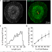
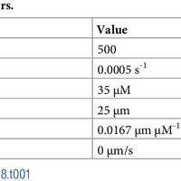
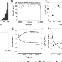
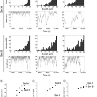
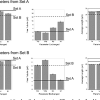
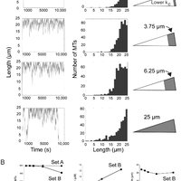
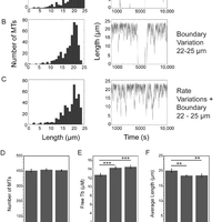
Microtubules are dynamic polymers required for a number of processes, including chromosome movement in mitosis. While regulators of microtubule dynamics have been well characterized, we lack a convenient way to predict how the measured dynamic parameters shape the entire microtubule system within a cell, or how the system responds when specific parameters change in response to internal or external signals. Here we describe a Monte Carlo model to simulate an array of dynamic microtubules from parameters including the cell radius, total tubulin concentration, microtubule nucleation rate from the centrosome, and plus end dynamic instability. The algorithm also allows dynamic instability or position of the cell edge to vary during the simulation. Outputs from simulations include free tubulin concentration, average microtubule lengths, length distributions, and individual length changes over time. Using this platform and reported parameters measured in interphase LLCPK1 epithelial cells, we predict that sequestering ~ 15–20% of total tubulin results in fewer microtubules, but promotes dynamic instability of those remaining. Simulations also predict that lowering nucleation rate will increase the stability and average length of the remaining microtubules. Allowing the position of the cell’s edge to vary over time changed the average length but not the number of microtubules and generated length distributions consistent with experimental measurements. Simulating the switch from interphase to prophase demonstrated that decreased rescue frequency at prophase is the critical factor needed to rapidly clear the cell of interphase microtubules prior to mitotic spindle assembly. Finally, consistent with several previous simulations, our results demonstrate that microtubule nucleation and dynamic instability in a confined space determines the partitioning of tubulin between monomer and polymer pools. The model and simulations will be useful for predicting changes to the entire microtubule array after modification to one or more parameters, including predicting the effects of tubulin-targeted chemotherapies.

Figures

  • Fig 1. MT length distributions for growing MT ends in LLCPK1 cells. (A) Cells were fixed 4 h after plating on a disc shaped adhesive surface. The position of the centrosome is shown in red and marked by the orange arrow. EB1-GFP marks growing MT plus ends. Lengths were measured assuming straight lines between the center of the centrosome and the distal tip of the EB1-GFP comet (B) Distribution of MT lengths averaged for 4 cells plated on a disc shaped pattern and having centered centrosomes.
  • Table 1. Common simulation parameters.
  • Table 2. Parameter sets for MT plus end dynamic instability.
  • Fig 2. Parameter Set A generates an array of long MTs, confined by the cell boundary. (A) Simulated MT length distribution for a radius of 25 μm. (B) Length of a single simulated MT over the 10,000 s of the simulation run. (C) The first 500 s of a simulation is shown to highlight the slower rate of MT polymerization as tubulin assembles into polymer (compare slopes at arrows). Free tubulin concentration declines rapidly during this time course (see graph in E). (D) Average MT length reaches a stable value of ~22 μm rapidly during simulations. (E,F) The number of MTs and free tubulin concentration, as well as their standard deviations, reach plateaus by ~ 10,000 s.
  • Fig 3. Comparison of outputs from parameter Sets A and B with varying total tubulin concentrations. (A) Simulation outputs as noted for parameter sets A and B and indicated total concentrations of tubulin. (B-D) Average ± sd values for MT number, free tubulin concentration and average MT length as a function of total tubulin concentration. Standard deviations are often smaller than the size of the data point.
  • Fig 4. Rescue frequency variations have the largest impact on MT number, length and free tubulin concentration.
  • Fig 5. Simulating two cytosolic zones differing in catastrophe frequency. Several groups have documented persistent MT growth (low catastrophe) in the cell interior. We simulated the outcomes of varying widths of an internal stable zone (16x lower catastrophe). (A) Simulations were run using parameter Set B. Data are plotted relative to the width of the peripheral, more dynamic (higher catastrophe) zone (shown in grey in the diagrams). The width of the internal, stable zone excludes MT plus ends because they quickly polymerize through this zone and confines dynamic instability to the more dynamic peripheral zone where catastrophes are much more likely. (B) As the peripheral, dynamic zone becomes narrower (lower values on X axis), Set B parameters predict output values similar to that generated by Set A.
  • Fig 6. Varying the position of the cell boundary shifts the length distribution, raises free tubulin concentration and shortens average MT length without changing MT number. The algorithm was modified to allow some parameters to vary randomly during a simulation. (A) Dynamic instability parameters were varied randomly by ± 1 sd around the means as described in the text. (B) The cell radius was reduced randomly by 0–3 μm to simulate small shifts in position of the cell boundary. (C) Combination of conditions in (A,B). See text for details. (D-F) The number of MTs is constant under the conditions shown in A-C, but free tubulin is slightly greater, and average length slightly shorter when the cell radius is reduced randomly by 0–3 μm during simulations. p< 0.0001, p = 0.001.

References Powered by Scopus

Decoding ALS: From genes to mechanism

1490Citations
N/AReaders
Get full text

Dynamic instability of individual microtubules analyzed by video light microscopy: rate constants and transition frequencies.

825Citations
N/AReaders
Get full text

Control of microtubule organization and dynamics: Two ends in the limelight

665Citations
N/AReaders
Get full text

Cited by Powered by Scopus

Autoregulation and repair in microtubule homeostasis

38Citations
N/AReaders
Get full text

Reorganization of paclitaxel-stabilized microtubule arrays at mitotic entry: roles of depolymerizing kinesins and severing proteins

27Citations
N/AReaders
Get full text

A self-organized synthetic morphogenic liposome responds with shape changes to local light cues

11Citations
N/AReaders
Get full text

Register to see more suggestions

Mendeley helps you to discover research relevant for your work.

Already have an account?

Cite

CITATION STYLE

APA

Cassimeris, L., Leung, J. C., & Odde, D. J. (2018). Monte Carlo simulations of microtubule arrays: The critical roles of rescue transitions, the cell boundary, and tubulin concentration in shaping microtubule distributions. PLoS ONE, 13(5). https://doi.org/10.1371/journal.pone.0197538

Readers over time

‘18‘19‘20‘22‘23‘24‘2505101520

Readers' Seniority

Tooltip

PhD / Post grad / Masters / Doc 15

63%

Researcher 7

29%

Professor / Associate Prof. 2

8%

Readers' Discipline

Tooltip

Biochemistry, Genetics and Molecular Bi... 11

50%

Agricultural and Biological Sciences 6

27%

Physics and Astronomy 3

14%

Computer Science 2

9%

Save time finding and organizing research with Mendeley

Sign up for free
0